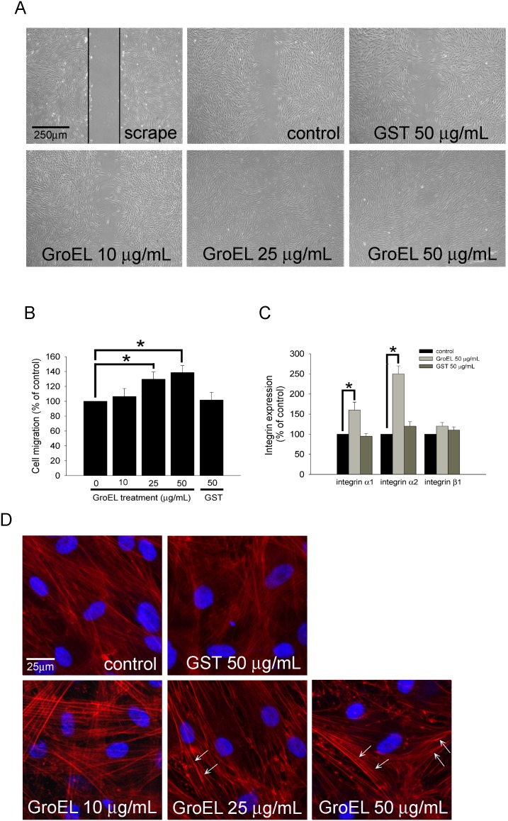
Figure 2. P. gingivalis GroEL increases PDL cell migration, possibly through activation of integrin α1 and α2 expression, as well as cytoskeletal reorganization.
(A) A wound-healing assay was used to evaluate the effect of GroEL on PDL cell migration. PDL cells were cultured with serum free medium (0 µg/mL; control), 10–50 µg/mL GroEL or 50 µg/mL GST for 24 h before wound scraping using a pipette tip. Images were taken 24 h after wound scraping. PDL cells migrating to the denuded area were counted based on the black base line. (B) PDL cells that migrated into the denuded area were quantified, and the magnitude of PDL cell migration was evaluated by counting the migrated cells in six random areas under high-power microscope fields (×100). (C) PDL cells were treated with serum-free medium (0 µg/mL; control), 50 µg/mL GroEL or 50 µg/mL GST for 12 h. The expression of integrin α1, α2, and β1 mRNA was analyzed using quantitative real-time PCR. Data are expressed as the mean ± SEM of three independent experiments and expressed as the percentage of control. *P<0.05 was considered significant. (D) F-actin was stained with rhodamine-phalloidin, and the staining was evaluated using confocal microscopy at 400× magnification. The visible parallel stress fibers are indicated as white arrows. DAPI was used to identify the nuclei of PDL cells.

