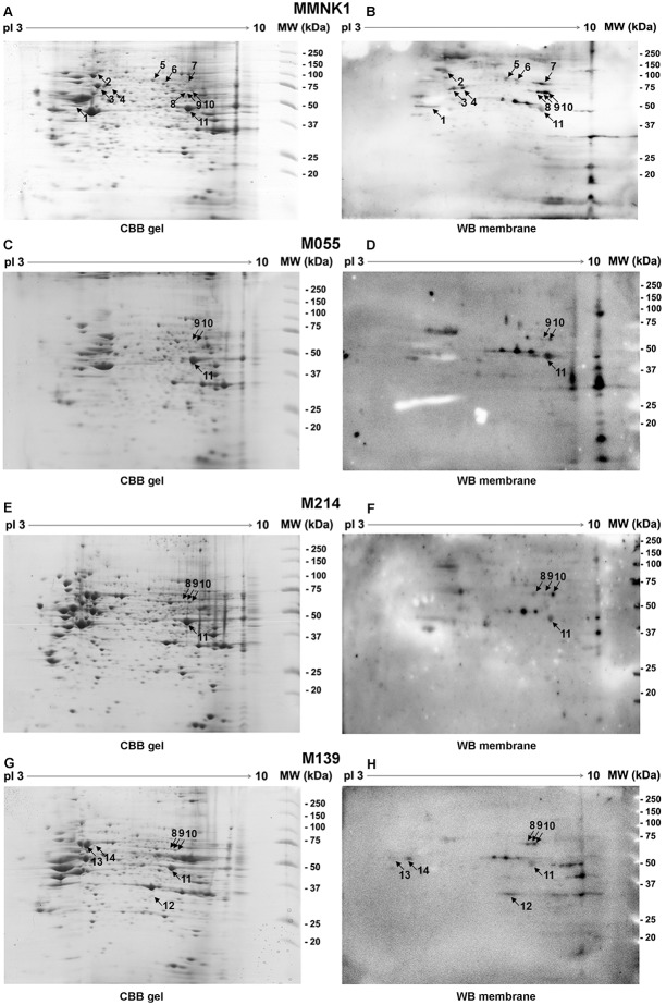
Figure 1. Two-dimensional (2D) gel electrophoresis and western blotting.
MMNK1 and CCA cell lines (M055, M214 and M139) lysates were separated by 2D electrophoresis and stained with coomassie brilliant blue (panels A, C, E and G). Parallel gels were transferred to PVDF and incubated with pooled plasma of patients with CCA (n = 10) and incubated with secondary anti-human IgG (panels B, D, F and H). Matching IgG autoantibodies spots were numbered 1–14 in both CBB gels and western blot membranes. Duplicate experiments were performed. Spot numbers 1, 11 and 13 are RNH1, ENO1 and HSP70, respectively.

