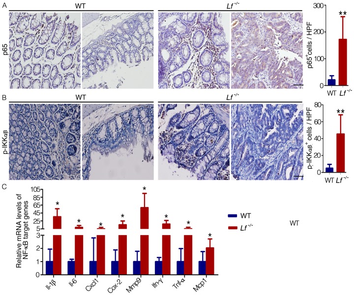
Figure 4. The canonical NF-.
κB pathway was activated in Lf −/− mice. The colons of AOM-DSS–treated mice were removed after 18 weeks. (A,B) Immunohistochemical analysis of nuclear p65 (A) and cytoplasmic p-IKKα/β (B) expression in the colon tissues. Cells that stained positive for p65 or for cytoplasmic p-IKKα/β were counted per high-powered field (HPF, 40× objective, bar = 50 µm). (C) mRNA expression levels of Il-1β, Il-6, Cxcl1, Cox-2, Mmp9, Ifn-γ, Tnf-α, and Mcp1 in the murine colons were assayed by qPCR. Each value represents the mean ± SD (n = 5 mice/group). The expression values for WT mice were set at 1 for each gene. *P<0.05, **P<0.01 versus WT mice.

