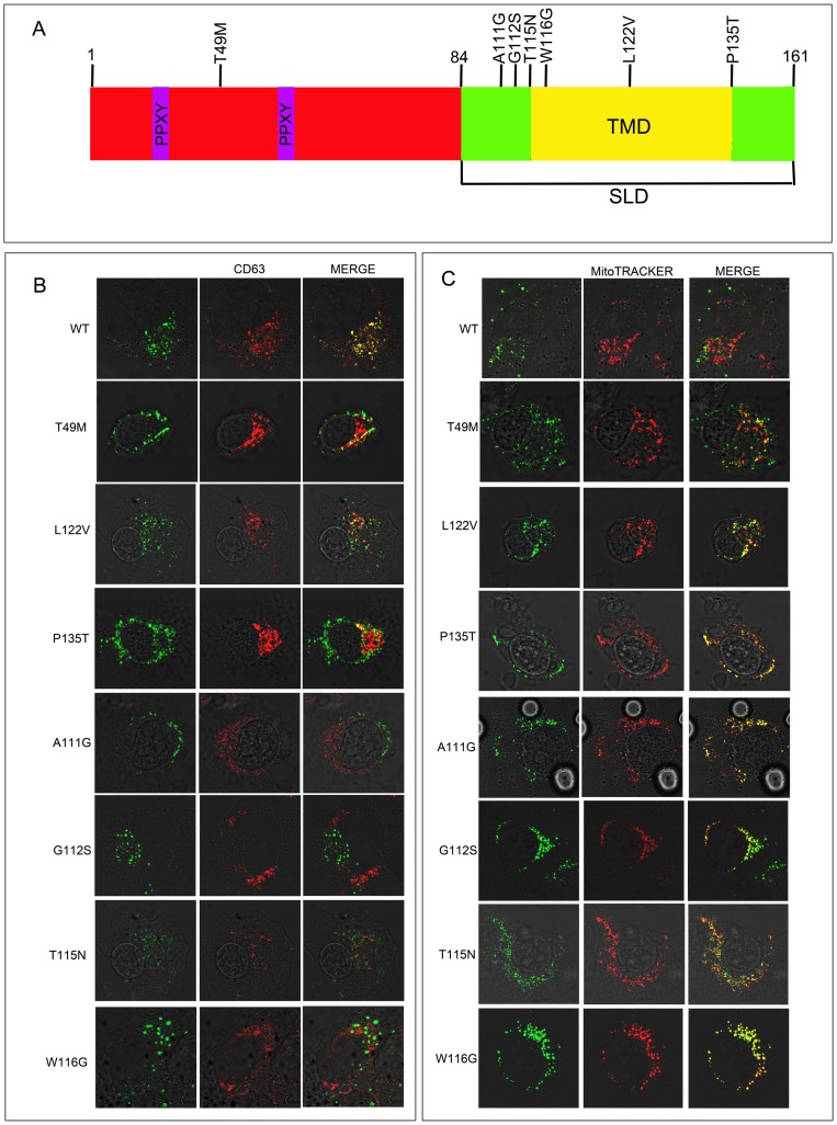
Figure 1. LITAF mutations associated with CMT disease.
(A) The position of mutations in LITAF that cause CMT1C are shown above the protein. The PPXY domains are shown in purple and the transmembrane domain (TMD) of the simple like domain (SLD) is shown in yellow. BGMK cells were transiently transfected with myc-tagged WT LITAF, T49M, A111G, G112S, T115N W116G, L122V or P135T. (B) Twenty-four hours post-transfection the cells were visualized using indirect immunofluorescence to detect LITAF using anti-myc antibodies (green) and the late endosome/lysosome was visualized using anti-CD63 (red) or (C) Twenty-four hours post-transfection the cells were incubated with MitoTRACKER Red FM for 2hrs. The cells were then fixed and permeabilized indirect immunofluorescence was performed to detect LITAF using anti-myc antibodies (green) and the mitochondria was visualized using MitoTRACKER Red FM. (red).

