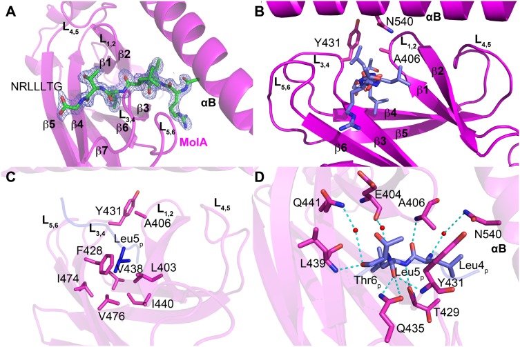
Figure 4. NRLLLTG peptide substrate binding by HSP70.
A. Omit electron density of the peptide substrate bound to the β subdomain of molecule A is contoured at 3.0 sigma. The NRLLLTG peptide is shown as a stick model in CPK coloring. B. Peptide binding site highlighting the flanking L1,2 and L3,4, and supporting L5,6 and L4,5 loops. The NRLLLTG peptide and arch residues are shown as stick models. C. Detailed view of the NRLLLTG-binding pocket in HSP70-NRLLLTG highlighting a network of van der Waals contacts with Leu5p of the NRLLLTG peptide. D. A close-up view of the interactions between Leu4p-Leu5p-Thr6p of the NRLLLTG peptide and the surrounding residues in HSP70-NRLLLTG. All interacting residues are shown as sticks and hydrogen bonds are shown as dotted lines.

