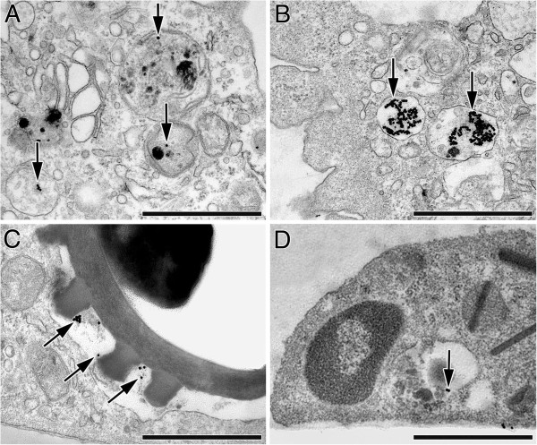
Figure 4.

Transmission electron micrographs of internalized AuNP (arrows) and spores in BAL cells. (A-C) AuNP in macrophages: (A) Single AuNP and small cluster in vesicles largely exceeding NP size and containing substances of mostly unknown origin. (B) Large AuNP clusters in vesicles slightly larger than the NP cluster. (C) AuNP co-localized with fungal spore in phagosome. (D) Small AuNP cluster in an eosinophil of an OVA-allergic mouse, in a vesicle exceeding NP size and containing other electron dense material of unknown origin. Bars: 1 μm.
