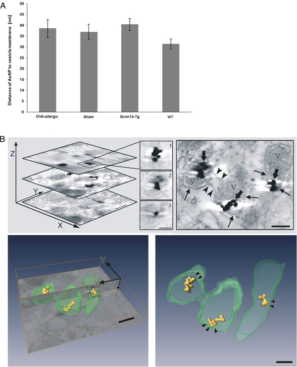
Figure 6.

AuNP localization with respect to the vesicle membrane. (A) Distance of AuNP to the vesicle membrane as measured on ultrathin sections of BAL macrophages (n = number of measurements). Data are expressed as mean values ± SEM per animal group. (B) Electron tomogram of small AuNP clusters in macrophage vesicle. Upper left: topmost, middle and lowest image of the 3D stack (bar: 100 nm) and magnified AuNP in different z-heights of the stack (1 - 3, bar: 50 nm). Upper right: middle image from the stack indicating connections between two vesicles (arrowheads) and showing AuNP (fat arrows) in close contact to vesicle membrane (arrows). Lower left: Bottom image of the tomogram stack (xy-plane) shows the sectioning xz-plane with automatic reconstruction of AuNP (yellow) and manual segmentation of vesicles (green). Lower right: reconstruction without the original electron micrograph. Arrowheads point to the space between NP and vesicle membrane. Bars of lower images: 100 nm.
