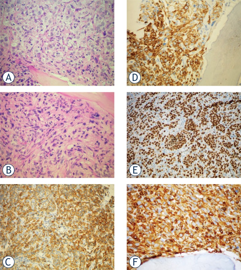
FIGURE 4.
A. Metastasis of the RCC in the bone: cells with copious clear cytoplasm and nuclei with prominent, eosinophilic nucleoli; bone trabecule is in the bottom part of the field; H&E 40x; B. More spindled tumor cells, »sacomatoid« differentiation; H&E, 40x; C. Positivity for CAM5.2; IHC CAM5.2, 20x; D. Positivity for RCC; IHC RCC, 40x; E. Positivity for PAX8, IHC PAX8, 20x; F. Positivity for Vimentin; IHC Vimentin, 40x.

