
Fig. 1.
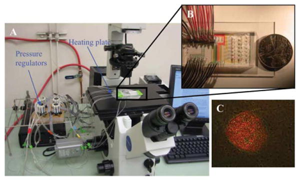
The setting of the microfluidic device for single hESC mRNA extraction. A. The system includes a microscope, a computer to control air pressure with pressure regulators, and a heating stage to heat the microfluidic chip to desire temperatures. B. A typical microfluidic chip. C. Merged image of immunofluorescent stained (Oct-3/4) and light microscope images from a pluripotent hESC colony. The hESC colony was labeled with mouse α human Oct-3/4 IgG and PE-conjugated rabbit anti-mouse IgG antibodies. Only cells in the center of the hESC colony expressed Oct-3/4. The intensity of the labeling indicates the Oct-3/4 positive cells expressed Oct-3/4 at different levels. The spontaneously differentiated cells around the colony do not express Oct-3/4.