
Fig. 2.
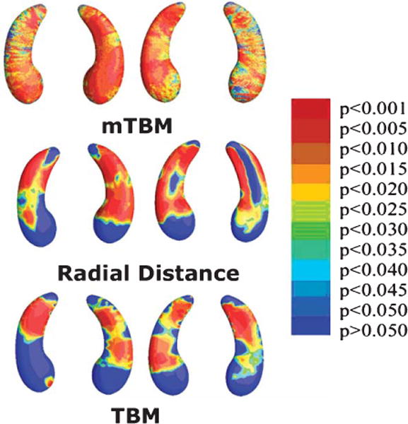
Mapping hippocampal correlates of tau proteins in the CSF. Here 3D maps show where alterations of hippocampal shape relate to levels of tau protein, measured in the CSF using lumbar puncture. To create these maps, we use a method called ‘multivariate tensor-based morphometry’ (mTBM; top row), as well as other methods to assess surface morphometry: radial distance maps (middle row) and standard TBM (bottom row). Non-blue colors show vertices with statistical differences, at the 0.05 level, uncorrected. Relationships were detected most sensitively with mTBM. Clearly, advanced mathematical methods can boost power to pick up associations between brain and CSF biomarkers, offering more detail than traditional measures of hippocampal volume. Adapted from Wang et al. [70] with permission of the authors and publishers.