Fig. 10.

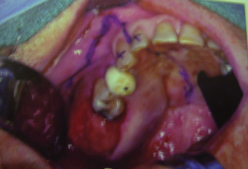
Case 3: Intraoral preoperative image showing the palatal bulge and a reddish pink soft compressible mass in the tuberosity region.

Case 3: Intraoral preoperative image showing the palatal bulge and a reddish pink soft compressible mass in the tuberosity region.