Fig. 11.

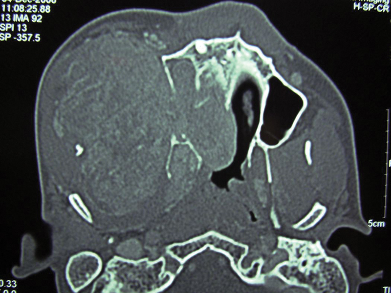
Case 3: Preirradiation axial section computed tomography demonstrating a huge parapharyngeal mass eroding the pterygoid plates and nasal septum and obliterating the nasal cavity and maxillary sinus on the same side extending onto the cheek.

Case 3: Preirradiation axial section computed tomography demonstrating a huge parapharyngeal mass eroding the pterygoid plates and nasal septum and obliterating the nasal cavity and maxillary sinus on the same side extending onto the cheek.