Fig. 13.

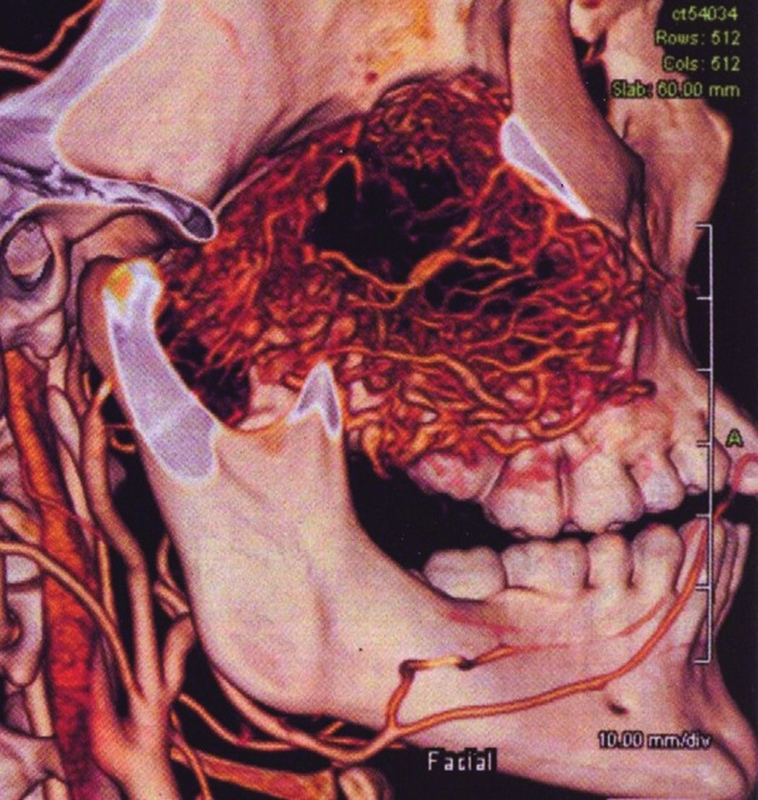
Case 3: Preoperative computed tomography angiography demonstrating the tumor with feeders from the ipsilateral internal maxillary, and the ascending pharyngeal and internal carotid arteries.

Case 3: Preoperative computed tomography angiography demonstrating the tumor with feeders from the ipsilateral internal maxillary, and the ascending pharyngeal and internal carotid arteries.