Fig. 19.

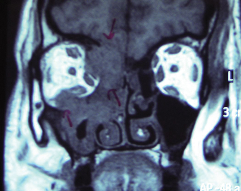
Case 7: Magnetic resonance imaging coronal section showing the lesion involving right nasal cavity, ethmoidal sinus, medial aspect, and floor of orbit, herniating into the right maxillary sinus.

Case 7: Magnetic resonance imaging coronal section showing the lesion involving right nasal cavity, ethmoidal sinus, medial aspect, and floor of orbit, herniating into the right maxillary sinus.