Fig. 25.

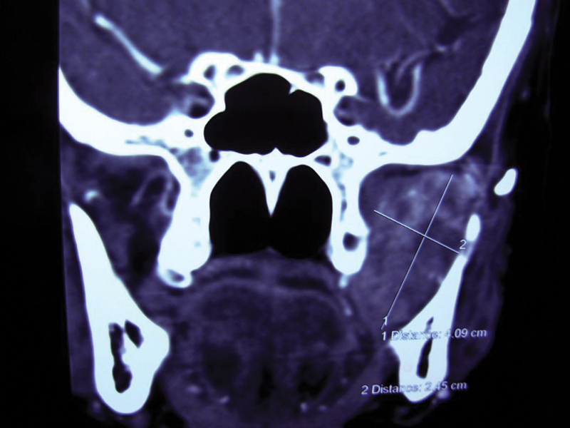
Case 9: Coronal computed tomography showing a well-defined 2 × 1 cm lesion in the left parapharyngeal area extending up to the infratemporal space.

Case 9: Coronal computed tomography showing a well-defined 2 × 1 cm lesion in the left parapharyngeal area extending up to the infratemporal space.