
Fig. 4.
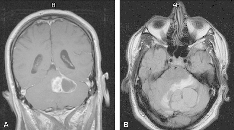
(A) Coronal T1-weighted postcontrast magnetic resonance imaging (MRI) and (B) axial flair MRI. Note the left cerebellar mass lesion with cystic areas. There is nonhomogeneous tumor enhancement (A) and some perilesional edema (B).
Official websites use .gov
A
.gov website belongs to an official
government organization in the United States.
Secure .gov websites use HTTPS
A lock (
) or https:// means you've safely
connected to the .gov website. Share sensitive
information only on official, secure websites.
(A) Coronal T1-weighted postcontrast magnetic resonance imaging (MRI) and (B) axial flair MRI. Note the left cerebellar mass lesion with cystic areas. There is nonhomogeneous tumor enhancement (A) and some perilesional edema (B).