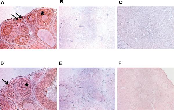
Figure 10.
Immunohistochemical analysis of Interleukin 16 and its receptor, CD4. Brown stain indicates presence of the specified protein. (A) Localization of IL16 in the ovary, particularly in granulosa cells of follicles (*). (B) Negative control experiment conducted without the use of primary antibody. (C) Negative control experiment conducted with non-specific Rabbit IgG. (D) Localization of CD4 receptor in the ovary, particularly in granulosa cells of follicles (*). (E) Negative control experiment conducted without the use of primary antibody. (F) Negative control experiment conducted with non-specific Goat IgG. Arrows point to developing primordial and primary follicles.

