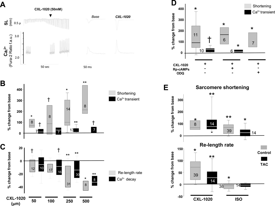
Figure 2. Influence of CXL-1020 on isolated cardiac myocytes from normal and failing heart.
A) (Left) Isolated myocyte sarcomere length (SL, upper tracing) and calcium transients (Ca2+, lower tracing) after exposure to CLX-1020. There is a marked rise in sarcomere shortening (SS) and a rise in the peak Ca2+ transient, as well as acceleration of the time for re-lengthening and calcium decline. B) Box-plots show percent change in SS and peak Ca2+ transient relative to baseline with incremental CXL-1020 dose. Only one dose was tested per myocyte; the sample size at each dose is provided in the plots. C) Percent reduction in sarcomere relengthening and calcium decay time. D) Percent change in SS and peak Ca2+ transient following CXL-1020 with or without co-inhibition of PKA (Rp-cAMPs) or soluble guanylate cyclase (ODQ). E) Percent change in SS and re-lengthening rate in myocytes isolated from control or failing hearts that are then exposed to either isoproterenol (ISO, 2.5 nM) or CXL-1020 (50 µM). † - p<0.05; * p<0.01; ** p≤0.001 versus respective (pre-CXL1020, or ISO) baseline, by Wilcoxan Sign-rank Test.

