FIGURE 5.
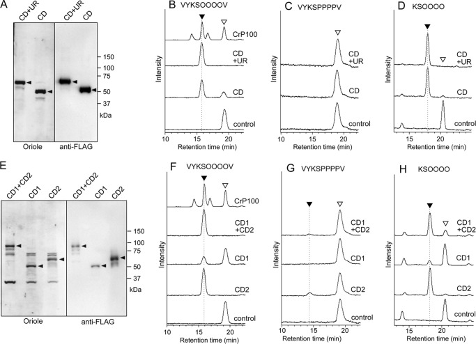
Expression of recombinant CrSGT1 and AtSGT1 and their enzymatic activity. A, recombinant CrSGT1 proteins were expressed by P. pastoris and then separated by SDS-PAGE. Proteins were stained using Oriole fluorescent gel stain (Bio-Rad). The anti-FLAG indicates Western detection of recombinant CrSGT1 proteins using an anti-FLAG tag antibody. Black arrowheads indicate recombinant CrSGT1 proteins. Expressed SGT regions (CD+UR and CD) are also shown in Fig. 4, A and B. B–D, recombinant CrSGT1 proteins (10 ng of CD+UR or CD) were incubated at 30 °C for 1 h with three types of acceptor peptides, and enzymatic products were analyzed by HPLC. The control was incubated with CD+UR treated at 95 °C for 10 min. Black and white arrowheads indicate enzymatic products of CrSGT1 and acceptor, respectively. The amino acid sequences of AtEXT, AtEXT-P, and AtEXTa peptides used as acceptors were, respectively, FITC-Ahx-VYKSOOOOV-NH2, FITC-Ahx-VYKSPPPPV-NH2, and FITC-GABA-KSOOOO-NH2. O indicates hydroxyproline. E, production of recombinant AtSGT1. Expressed recombinant proteins were separated by SDS-PAGE, and proteins were stained with Oriole fluorescent gel stain or subjected to immunoblot analysis. Recombinant AtSGT1 proteins were detected using an anti-FLAG tag antibody. Black arrowheads indicate recombinant AtSGT1 proteins. Expressed SGT regions (CD1+CD2, CD1, CD2) are also shown in Fig. 4, A and B. F–H, AtSGT1 proteins (200 ng of CD1+CD2, CD1, and CD2) expressed by P. pastoris were incubated at 30 °C for 12 h with three acceptor peptides, and enzymatic products were analyzed by HPLC. The control was incubated with CD1+CD2 treated at 95 °C for 10 min. Black and white arrowheads are same as B–D. B and F, a 10-μg P100 fraction of C. reinhardtii CC-503 was also used for galactosyltransferase activity assay as shown in Fig. 1A, which is indicated as CrP100.

