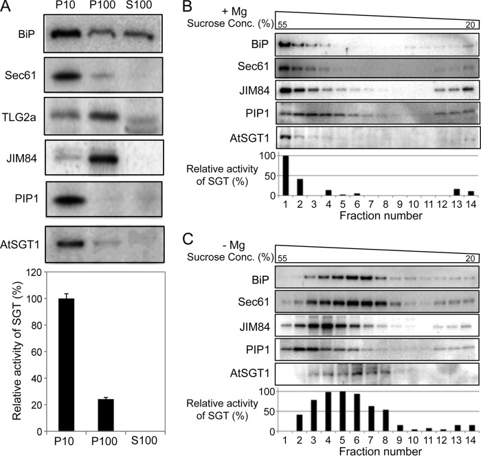
FIGURE 9.
Subcellular localization of AtSGT1. A, subcellular fractionation of AtSGT1. P10, P100, and S100 fractions were prepared from Col-0 cells, and AtSGT1 and various markers were detected by immunoblotting with specific antibodies that recognize BiP or Sec61 (for the ER membrane), TLG2a or JIM84 (Golgi apparatus), PIP1 (plasma membrane), and AtSGT1. SGT activity in each fraction was also shown. 100% corresponds to 9.5 × 10⁻³ unit (nmol/min/mg protein) incubated with P10 fraction extracted from Col-0 for 10 h. B and C, total protein extract (excluding cell debris) was prepared from Col-0 cells in buffers containing either Mg2+ (+Mg) or EDTA (−Mg). After sucrose density gradient centrifugation, the sample was separated into 14 fractions. The presence of AtSGT1 in these fractions was determined by immunoblotting and measurement of SGT activity. The distribution of marker proteins was analyzed by immunoblotting. 100% corresponds to 14.8 × 10⁻⁶ units and 26.8 × 10⁻⁶ units (nmol/min/fraction) incubated with fraction 1 in B and fraction 5 in C for 10 h, respectively.

