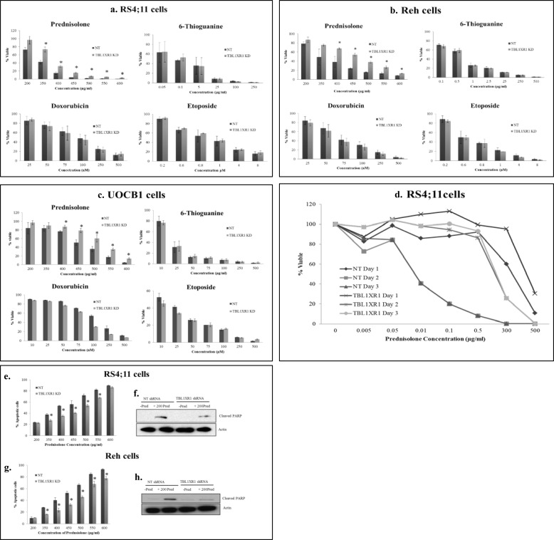
FIGURE 4.
Nontargeting shRNA (NT) and TBL1XR1 targeting shRNA cells were treated with increasing concentrations of prednisolone, doxorubicin, and etoposide for 24 h or thioguanine for 48 h. a–d, cell viability was measured by Cell Titer Glo assay. a–d, RS4;11 cells (a), Reh cells (b), UOCB1 cells (c), RS4;11 control (d, NT), and TBL1XR1 knockdown lines were treated with prednisolone at indicated concentrations for 24, 48, or 72 h, and then cell viability was determined by Cell Titer Glo assay. e and g, Nontargeting shRNA (NT) and TBL1XR1 targeting shRNAs were treated with prednisolone for 24 h, and levels of apoptosis were determined by flow cytometry RS4;11 cells (e) and Reh cells (g). f and h, levels of apoptosis were confirmed by Western blot for downstream apoptotic marker cleaved PARP in NT cells, as well as TBL1XR1 knockdown cells in the presence or absence of 200 μg/ml of prednisolone RS4;11 cells (f) and Reh cells (h). Error bars represent standard deviations of two or three replicate experiments. *, significant change with a p value less than or equal to 0.05.

