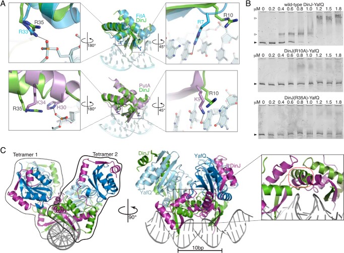
FIGURE 4.
DNA recognition by the N terminus of DinJ at the dinJ promoter. A, modeling of DinJ binding to DNA based on the structure of FitA-DNA complex (blue, PDB code 2BSQ) and PutA-DNA complex (purple, PDB code 2RBF). PutA and FitA residues required for DNA binding as well as structurally equivalent DinJ residues are displayed as sticks. B, EMSAs show that the wild-type DinJ-YafQ complex binds to its promoter, whereas DinJ(R10A)-YafQ and DinJ(R35A)-YafQ fail to bind DNA. Black-filled arrows and open arrows indicate unbound and TA-bound DNA, respectively. C, model of YafQ-(DinJ)2-YafQ bound to two inverted DNA repeats (DNA shown in gray). A minor steric clash occurs between DinJ α1 (magenta) and DinJ′ α1 (green) (outlined in red; zoomed in, right panel).

