
FIGURE 3.
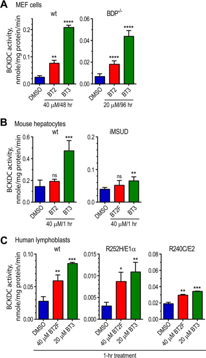
BT2, BT2F, and BT3 significantly increase BCKDC activity in wild-type and MSUD cells. A, wild-type MEF cells were cultured with 40 μm BT2, BT2F, or BT3 for 48 h with DMSO as a control. BDP−/− cells manifesting intermediate MSUD were grown in the presence of 20 μm BT2 or BT3 for 96 h. BCKDC activity was measured by the intact cell assay using α-keto[1-14C] isovalerate as a substrate. B, primary hepatocytes prepared from wild-type MSUD or iMSUD mice were incubated in the Krebs buffer with BT2 or BT3 (40 μm) for 1 h and assayed for BCKDC activity in a reconstituted reaction mixture. C, harvested lymphoblasts from control subjects and intermediate MSUD patients carrying R252H/E1α or R240C/E2 (both mature sequences) were incubated in the Krebs buffer for 1 h with 40 μm BT2 or BT2F or 20 μm BT3. Wild-type and mutant BCKDC activity was measured by the intact cell assay. Error bars, S.D.