
FIGURE 6.
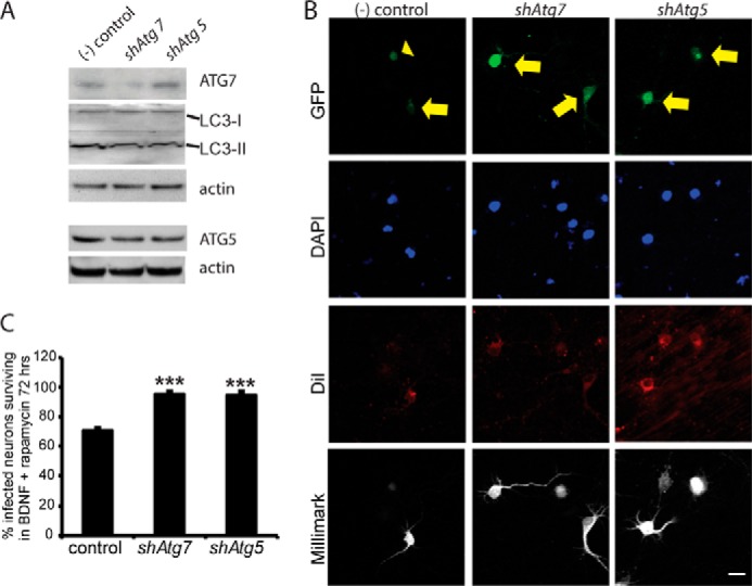
Autophagy reduction rescues neurons co-treated with BDNF and rapamycin. A, Western blots of lentivirus-infected cells show Atg7 and Atg5 knockdown by shAtg7 and shAtg5, respectively. Actin normalization revealed specific knockdown by each shRNA construct. Knockdown effects were more evident in survival experiments, in which only GFP+ neurons were analyzed, than in Western blot validation, probably reflecting low transfection efficiency. Functional reduction in autophagy is shown by reduced ratio of LC3-II to LC3-I proteins. B, infected cells express GFP and were scored as living if they stained positive for DiI and for the pan-neuronal marker Millimark. Infected cells were scored as dead if they expressed GFP but were DiI-negative and stained negative or at a low level with Millimark. C, quantification of B, three independent experiments for a total of at least 75 transfected neurons per group. Autophagy suppression increases neuron survival in co-treatment with BDNF and rapamycin. Scale bar, 20 μm, ***, p < 0.001.