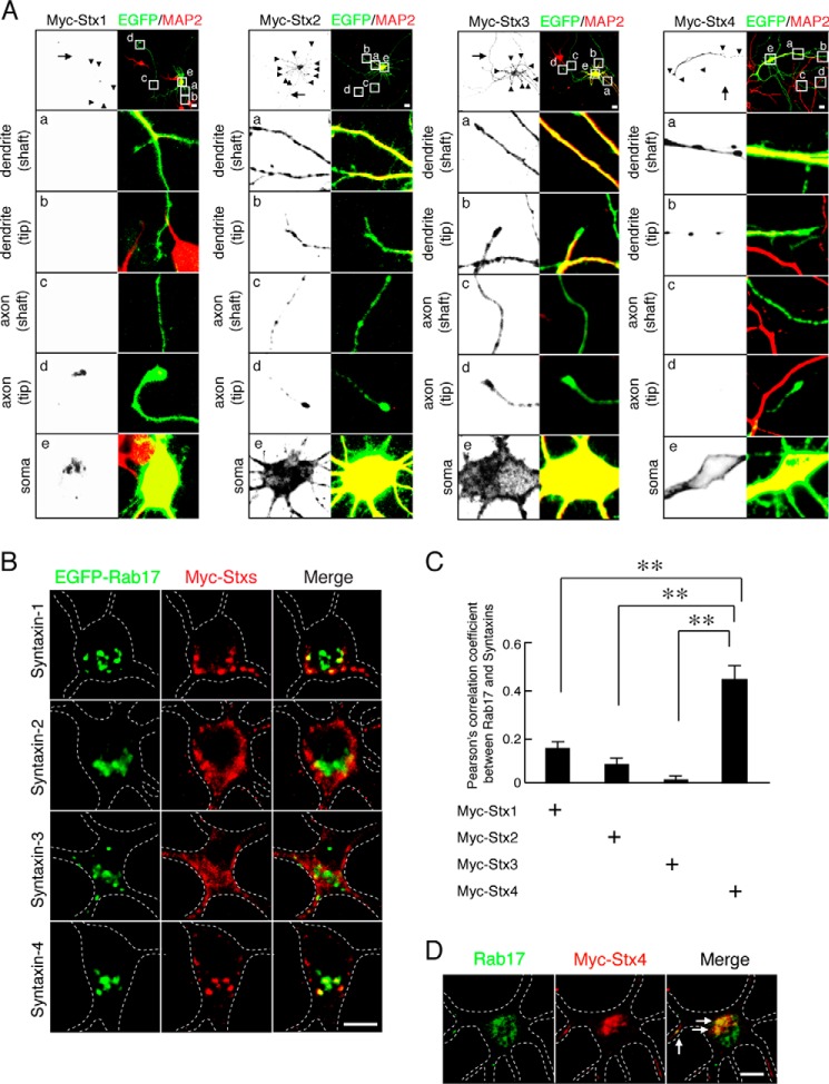
FIGURE 3.
Rab17 co-localizes with Syntaxin-4 in the soma. A, representative images of plasma membrane-associated Syntaxins in developing neurons. At 8 DIV, mouse hippocampal neurons were transfected with pCMV-Myc-Stx1 (left panel), pCMV-Myc-Stx2 (2nd panel from the left), pCMV-Myc-Stx3 (3rd panel from the left), or pCMV-Myc-Stx4 (right panel), and at 11 DIV, the neurons were fixed and subjected to immunocytochemistry with antibodies against Myc (black) and MAP2 (red). The arrows and arrowheads point to axons and dendrites, respectively. The lower four panels a–d are magnified views of the boxed areas in the top right panels. Bar, 10 μm. B, representative images of Rab17 and plasma membrane-associated Syntaxins at the soma in developing neurons. At 8 DIV, mouse hippocampal neurons were transfected with pEGFP-Rab17 together with pCMV-Myc-Stx1 (top panel), pCMV-Myc-Stx2 (2nd panel), pCMV-Myc-Stx3 (3rd panel), or pCMV-Myc-Stx4 (bottom panel), and at 11 DIV, the neurons were fixed and subjected to immunocytochemistry with antibodies against Myc (red) and MAP2. The dashed lines indicate dendritic shafts identified as MAP2-positive areas. Bar, 5 μm. C, quantification of the Pearson's correlation coefficient between EGFP-Rab17 and Myc-Syntaxin-1 (n = 10), Myc-Syntaxin-2 (n = 10), Myc-Syntaxin-3 (n = 10), and Myc-Syntaxin-4 (n = 10), as shown in A. **, p < 0.0025. D, representative images of endogenous Rab17 and Myc-Syntaxin-4 in the soma at developing neurons. At 8 DIV, hippocampal neurons were transfected with pCMV-Myc-Stx4, and at 11 DIV, the neurons were fixed and subjected to immunocytochemistry with antibodies against Rab17 (green), Myc (red), and MAP2. The dashed lines indicate MAP2-positive areas, and the arrows indicate the co-localization points. Bar, 5 μm.

