FIGURE 6.
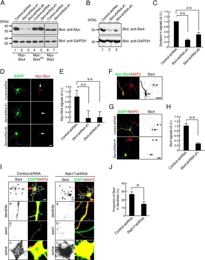
Rab17 is required for dendritic trafficking of endogenous Syntaxin-4. A, HEK293T cells were transfected with pFIV-Control (lanes 1, 4, and 6), pFIV-shStx4-1 (lanes 2, 5, and 7), or pFIV-shStx4-2 (lane 3) together with pCMV-Myc-ratStx4 (lanes 1–3), pCMV-Myc-Stx4SR (lanes 4 and 5), or pCMV-Myc-Stx-3 (lanes 6 and 7). Two days after transfection, the cells were lysed and subjected to immunoblot analysis with anti-Myc antibody (upper panel) and anti-GAPDH antibody (lower panel). B, Neuro2A cells were transfected with pFIV-Control (lane 1), pFIV-shStx4-1 (lane 2), or pFIV-shStx4-2 (lane 3). Two days after transfection the cells were lysed and subjected to immunoblot analysis with anti-Syntaxin-4 antibody (upper panel) and anti-GAPDH antibody (lower panel). C, quantification of endogenous Syntaxin-4 of control-shRNA, Stx4-shRNA-1 and Stx4-shRNA-2 as shown in B. A.U., arbitrary units. **, p < 0.0025. D, representative images of Myc-Syntaxin-4-expressing neurons in the presence and absence of Stx4-shRNA. At 8 DIV, rat hippocampal neurons were transfected with pCMV-Myc-ratStx4 together with pFIV-Control, pFIV-shStx4-1, or pFIV-shStx4-2, and at 11 DIV, the neurons were fixed. The neurons were stained by Myc (red). Bar, 10 μm. E, quantification of the Myc-Syntaxin-4 of control-shRNA-transfected neurons (n = 10), Stx4-shRNA-1-transfected neurons (n = 10), and Stx4-shRNA-2-transfected neurons (n = 10) as shown in B. A.U., arbitrary units. **, p < 0.0025. F, representative images of Myc-Syntaxin-4-expressing neurons stained by Syntaxin-4 antibody. At 8 DIV, rat hippocampal neurons were transfected with pCMV-Myc-Stx4, and at 11 DIV, the neurons were fixed. The neurons were stained by Myc (green), Syntaxin-4 (black), and MAP2 (red). Bar, 10 μm. G, representative images of endogenous Syntaxin-4 in the presence and absence of Stx4-shRNA. At 8 DIV, rat hippocampal neurons were transfected with pFIV-Control and pFIV-shStx4-1, and at 11 DIV, the neurons were fixed. The neurons were stained by Syntaxin-4 (black) and MAP2 (red). Bar, 10 μm. H, quantification of endogenous Syntaxin-4 of control-shRNA-transfected neurons (n = 10) and Stx4-shRNA-1-transfected neurons (n = 10) shown in G. A.U., arbitrary units. **, p < 0.0025. I, representative images of endogenous Syntaxin-4 in the Rab17-shRNA-transfected neurons. At 8 DIV, mouse hippocampal neurons were transfected with pSilencer-CMV-EGFP-Control or pSilencer-CMV-EGFP-shRab17, and at 11 DIV, the neurons were fixed and subjected to immunocytochemistry with antibodies against Syntaxin-4 (black), GFP (green), and MAP2 (red). The arrows and arrowheads point to axons and dendrites, respectively. The lower three panels a–c are magnified views of the boxed areas in the top right panels. Bar, 10 μm. J, quantification of the proportion of endogenous Syntaxin-4 in the control neurons (n = 10) and Rab17-shRNA-transfected neurons (n = 10) at the dendrites, shown in I. The rate of translocated level of Syntaxin-4 was calculated by dividing the dendrite Syntaxin-4 fluorescence intensity by the total Syntaxin-4 fluorescence intensity. *, p < 0.025.

