FIGURE 8.
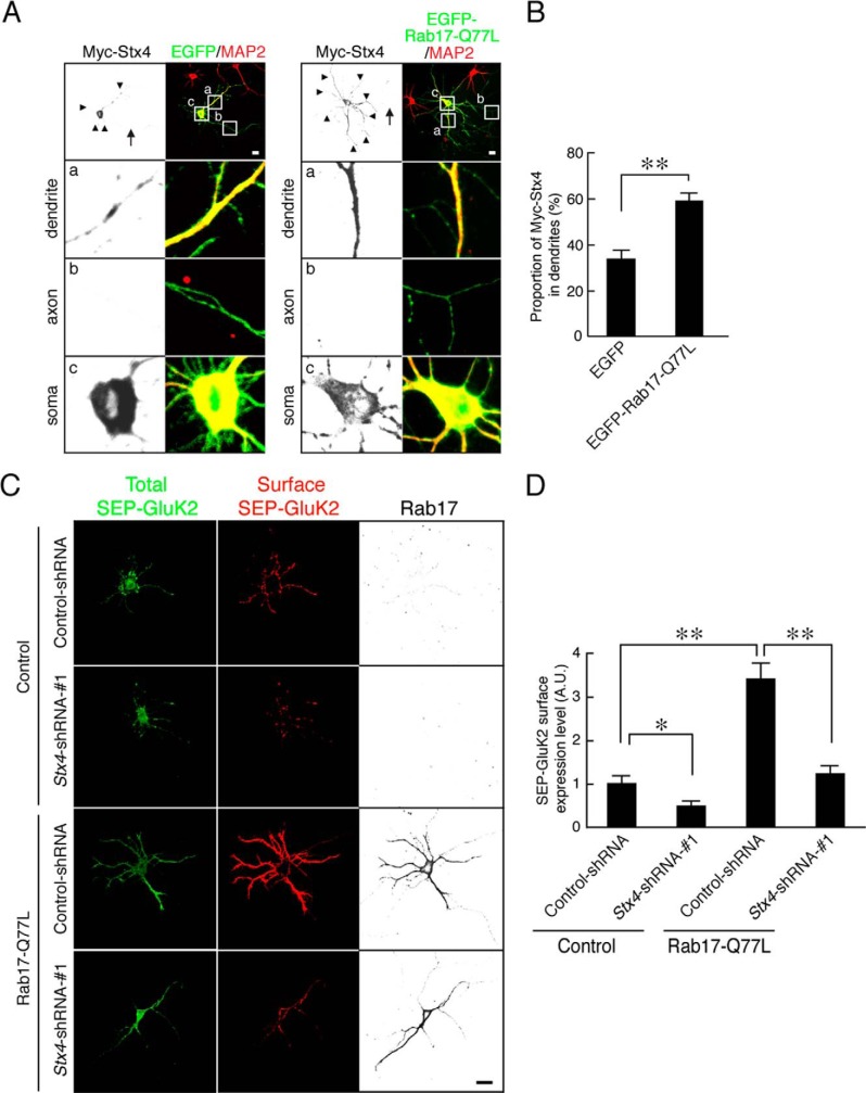
Active form of Rab17 promotes surface expression of GluK2 by enhancing Syntaxin-4 translocation to dendrites. A, representative images of Myc-Syntaxin-4 in active form Rab17-expressing neurons. At 8 DIV, rat hippocampal neurons were transfected with pCMV-Myc-Stx4 together with pEGFP-C2 or pEGFP-Rab17-Q77L. At 11 DIV, the neurons were fixed and subjected to immunocytochemistry with antibodies against Myc (black) and MAP2 (green). The bottom three panels a–c are magnified views of the boxed areas in the top right panels. Bar, 10 μm. B, quantification of the proportion of Myc-Syntaxin-4 in the dendrites in the presence of EGFP (n = 10) and EGFP-Rab17-Q77L as shown in A. The rate of translocated level of Myc-Syntaxin-4 was calculated by dividing the dendrite Myc-Syntaxin-4 fluorescence intensity by the total Myc-Syntaxin-4 fluorescence intensity. **, p < 0.0025. C, representative images of surface expression level of SEP-GluK2 in active form Rab17 and Stx4-shRNA-transfected neurons. At 8 DIV, rat hippocampal neurons were transfected with pCDNA3-SEP-Myc-GluK2 together with pCAG-Control and pFIV-Control, pCAG-Control, and pFIV-shStx4-1, pCAG-Myc-Rab17-Q77L, and pFIV-Control, or pCAG-Myc-Rab17-Q77L and pFIV-shStx4-1. At 11 DIV, the neurons were fixed and subjected to immunocytochemistry with antibodies against GFP (surface, red and total, green) and Rab17 (black). Bar, 10 μm. D, quantification of the surface expression level of SEP-GluK2 of control neurons (n = 10), Stx4-shRNA-1-transfected neurons (n = 10), Rab17-Q77L-transfected neurons (n = 10), and Rab17-Q77L- and Stx4-shRNA-1-transfected neurons (n = 10) as shown in C. Surface expression level of SEP-GluK2 was calculated by dividing the surface SEP-GluK2 fluorescence intensity by the total SEP-GluK2 fluorescence intensity. A.U., arbitrary units. *, p < 0.025; **, p < 0.0025.

