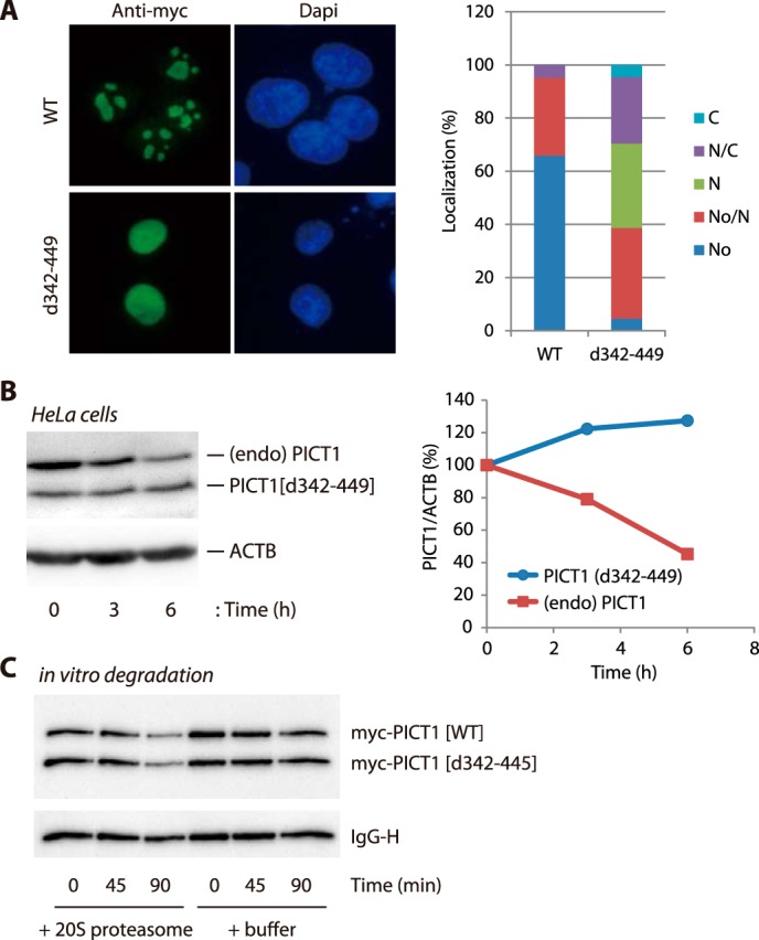
FIGURE 7.

PICT1 deletion mutant (d342–449) is resistant to nucleolar stress-induced degradation and shows nuclear localization. A, H1299 cells were transfected with Myc-PICT1/pEF1 (WT) or Myc-PICT1(d342–449)/pEF1 (d342–449). After a 2-day incubation, intracellular localization of ectopically expressed Myc-tagged PICT1 protein was analyzed by immunofluorescence assay as described under “Experimental Procedures.” One hundred cells were analyzed for each sample, and localization patterns of Myc-tagged PICT1 are presented in the right panel. No, nucleolar; No/N, nucleolar and nuclear; N, nuclear; N/C, nuclear and cytosolic; C, cytosolic. B, H1299/Myc-PICT1(d342–449) cells were treated with 10 ng/ml doxycycline for 24 h to induce expression of PICT1(d342–449) mutant protein in cells. The cells were treated with 1 mm FUrd for the indicated times, and cell lysates were prepared and subjected to immunoblot analysis as described under “Experimental Procedures.” The calculated PICT1/ACTB value of each control (0 h) was set to 100%, and normalized values are presented in the right panel. PICT1(d342–449), blue; endogenous PICT1, red. C, comparable amount of immunopurified Myc-PICT1 proteins (WT and d342–449) were mixed and subjected to 20 S proteasome-mediated in vitro degradation assay. After incubation for the indicated times, Myc-tagged proteins and mouse IgG heavy chain (IgG-H) was detected by immunoblot analysis as described under “Experimental Procedures.”
