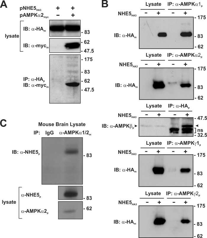
FIGURE 3.
NHE5 forms a macromolecular complex with AMPK subunits in heterologous and native tissue. A, AP-1 cells stably expressing triple HA-tagged NHE5 (NHE5HE3) were transiently transfected (24 h) with empty vector or full-length Myc-tagged AMPKα2 (AMPKα2myc). Cell lysates were prepared and incubated with a rabbit polyclonal anti-HA antibody (α-HAp) to immunoprecipitate NHE5HA3. The cell lysates and immunoprecipitates (IP) were analyzed by SDS-PAGE and immunoblotted (IB) with either a monoclonal anti-HA antibody (α-HAm) or anti-Myc (α-mycm) to detect NHE5HA3 or AMPKα2myc, respectively. B, to detect association of endogenous AMPK subunits with NHE5HA3, cell lysates were prepared from untransfected AP-1 cells or AP-1 cells stably expressing NHE5HA3 and then incubated with rabbit polyclonal antibodies against endogenous AMPKα1, -α2, -γ1, and -γ2 subunits. To detect a complex between NHE5 and the native AMPK β1/2 subunits, cell lysates were incubated with a rabbit polyclonal anti-HA antibody. The immunoprecipitates were analyzed by SDS-PAGE and immunoblotting with either mouse monoclonal anti-HA antibody to detect NHE5HA3 or with a commercially available pan anti-AMPKβ antibody that recognizes both the β1 and β2 subunits. C, to detect association of native AMPKα subunits with NHE5 in brain tissue, cell lysates were prepared from mouse brain (postnatal day 3) and then incubated with a control mouse IgG antibody or a mouse monoclonal antibody that recognizes both AMPKα1 and -α2 (α-AMPKα1/2m). The immunoprecipitates and cell lysates were analyzed by SDS-PAGE and immunoblotting. The IP blot was probed with a rabbit polyclonal antibody that specifically detects NHE5 (α-NHE5p). The lysate blots were probed with polyclonal antibodies that recognize NHE5 or AMPKα2. Data are representative of three independent experiments.

