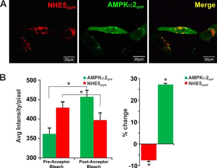
FIGURE 4.

NHE5 and AMPKα2 colocalize in transfected AP-1 cells. A, immunofluorescence confocal microscopy images of AP-1 cells transiently co-transfected (48 h) with monomeric cherry fluorescent protein-tagged NHE5 (NHE5ChFP, red) and enhanced green fluorescent protein-tagged AMPKα2 (AMPKα2GFP, green). Overlapping signals in merged image are yellow. B, fluorescence (or Forster) FRET analysis of NHE5ChFP and AMPKα2GFP interactions were determined by the acceptor photobleaching method (see ”Experimental Procedures“). Photobleaching of NHE5ChFP resulted in an increase in the GFP signal previously transferred to ChFP during FRET (left panel). Percent change in average fluorescence per pixel, relative to pre-bleaching of NHE5ChFP is shown in the right panel. Data are representative of at least three independent experiments. Asterisks indicate p < 0.05, Student's paired t test.
