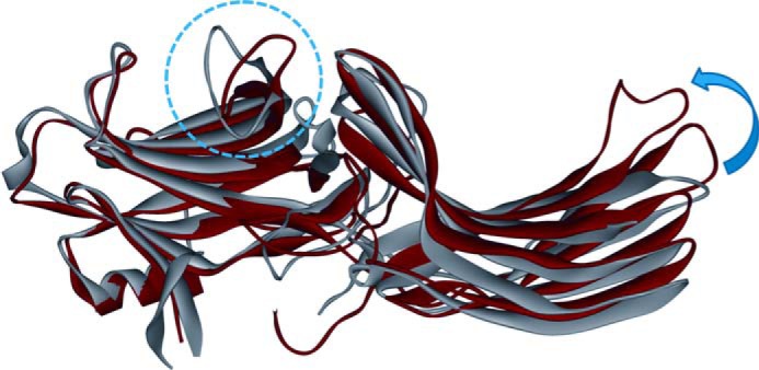
FIGURE 7.

The crystal structures of inactive arrestin-2 (PDB entry 1G4M) and V2Rpp-bound arrestin-2 (PDB entry 4JQI) overlaid by alignment of the N domains. The inactive structure is shown in gray, and the phosphorylated receptor peptide-bound structure is shown in red. The conformational change of the finger loop is highlighted by the blue dotted circle. The interdomain rotation observed in the crystal structure is indicated by the blue arrow.
