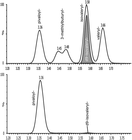
Fig. 1.

Chromatographic separation of the C5 isoforms (solved in methanol/water). The deuterated d9 isovalerylcarnitine (internal standard) is shown in the bottom chromatogram. Sample of the patient (day 3) eluted from the dried blood spot (bottom chromatogram). The pivaloylcarnitine was the most prominent peak, all other C5 acylcarnitines were only detected in low concentrations
