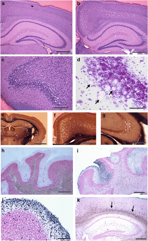
Fig. 2.

Examples of pathology in selected locations in HE-stained sections (a–d), Weil-myelin-stained sections (e–g), and amino cupric silver–stained sections (h-k). Extensive vacuolation is present in hippocampus CA3, retrosplenial cortex, and cerebral cortex layer 5 in an affected mouse (b) compared to a control mouse (a). Vacuolation is prominent in piriform area in frontal lobe (c). Enlargement of vacuoles in part of hippocampus CA3 in Nissl stain reveals intracytoplasmic vacuoles; arrows indicate cells where a flattened nucleus is visible adjacent to a vacuole (d). Low-magnification view of one hemisphere (e) demonstrates restricted locations of vacuoles in cortex and hippocampus, with higher magnification views, indicated by rectangles, showing vacuolation in retrosplenial cortex and cerebral cortex layer 5 (f) and part of hippocampus CA3 (g). The absence of cupric silver reaction product in control cerebellum molecular layer, except for red blood cells in vessels (h) contrasts with reaction product in a section of cerebellum from an affected mouse showing degeneration staining in the molecular layer, variable among folia (i). Large numbers of silver-stained puncta consistent with degenerating nerve terminals were observed in some folia (j). Localized degeneration staining was consistently observed in deep layers of cerebral cortex of affected mice (k). Scale bars and mouse ID numbers for each image: a-300 μm, #352; b-300 μm, #262; c-250 μm, #264; d-20 μm, #349; e- 900 μm, #348; f – 275 μm, #348; g-200 μm, #349; h-520 μm, #352; i-520 μm, #348; j-250 μm, #349; k-420 μm, #262
