
Figure 2. Sequence logos representing the recognition specificity of the SH2 domain family.
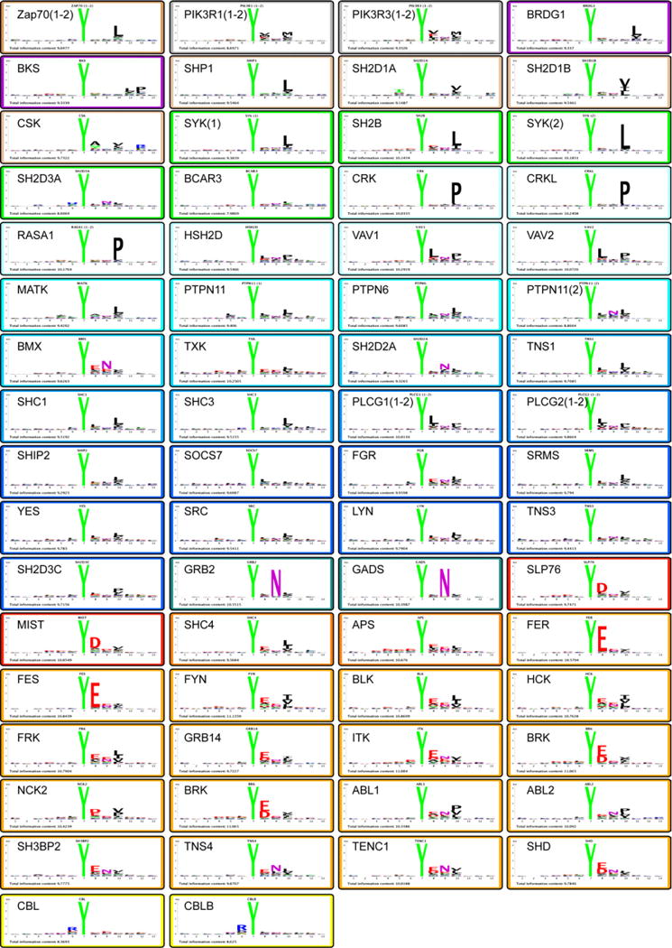
For each SH2 domain, the peptides whose binding signal was higher than the average signal plus two standard deviations were aligned on the phosphorylated tyrosine. These peptides were used to draw the peptide logos by a Logo drawing tool implemented in the PepSpot database (see Extended Results in Supplementary materials). Domain Logos of the same specificity class are framed in identical colors. The Logo total information content is also indicated in each frame (See also Supplementary Table S2).