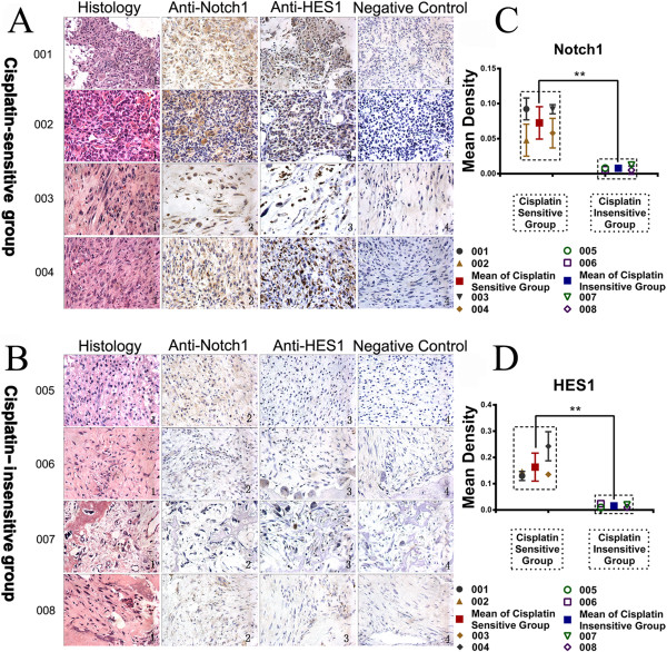
Figure 1.

Immunohistochemistry showed the expression of Notch1 and HES1 in cisplatin sensitive group and insensitive group. The histopathology revealed the irregular nuclei, some spindle cells, and even destroyed bone (A and B, Column 1). Immunohistochemical examination of Notch1 and HES1 were carried out in different patients. There is significant more expression of Notch1 and HES1 in cisplatin sensitive patients than cisplatin insensitive patients in gross appearance (A and B, Column 2 and 3) and semi-quantitative scatter plot below (C and D). Column 4 in A and B was the negative control with the secondary antibody alone without primary antibody. (**P < 0.01).
