
Figure 2.
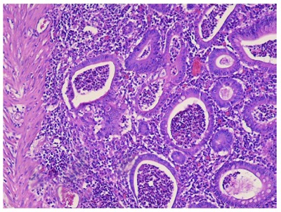
Histopathological image from the excised colon, typical of ulcerative colitis. This image demonstrates marked lymphocytic infiltration (blue/purple) of the intestinal mucosa and architectural distortion of the crypts (right side of the image). The inflammation is shallow and affects only the mucosa sparing the muscularis mucosal (left side).