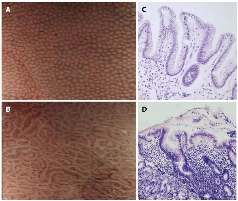
Figure 2.

Narrow band imaging of the gastric body. A: Normal mucosa shows uniform small, round pits surrounded by honeycomb-like subepithelial capillary networks (SECNs); B: Helicobacter pylori (H. pylori)-associated gastritis shows obviously enlarged, oval or prolonged pits; C: Corresponding histologic features of Figure 2A; D: Corresponding histologic features of Figure 2B.
