
Figure 6.
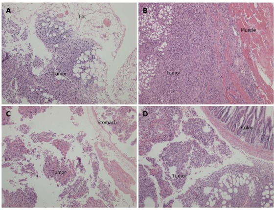
Cross-sections and hematoxylin-eosin staining of intra-abdominal disseminated foci observed under light microscopy (× 100). A: Tumor implanted in the fat tissue; B: Tumor implanted in the abdominal wall; C: Tumor implanted in the stomach wall; D: Tumor implanted in the colon serosal.