
Figure 4.
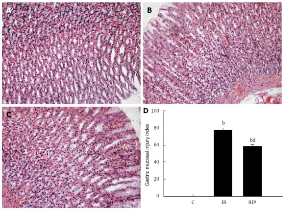
Histologic evaluation of gastric tissue. Hematoxylin and eosin staining of gastric sections obtained from A: Sham surgery; B: Ischemia reperfusion (IR); C: Remote ischemic postconditioning (RIP) after 6 h (magnification × 200); D: Quantitative scores for acute gastric lesions from sham, IR and RIP groups (mean ± SD; n = 6); bP < 0.01 vs sham group; dP < 0.01 vs IR group.