
Figure 1.
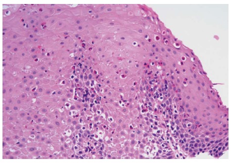
Reactive squamous mucosa with marked increased intraepithelial eosinophilia involving the entire thickness of the epithelium, occasional eosinophilic microabscesses and degranulation of eosinophils (HE stain, × 400).
Official websites use .gov
A
.gov website belongs to an official
government organization in the United States.
Secure .gov websites use HTTPS
A lock (
) or https:// means you've safely
connected to the .gov website. Share sensitive
information only on official, secure websites.
Reactive squamous mucosa with marked increased intraepithelial eosinophilia involving the entire thickness of the epithelium, occasional eosinophilic microabscesses and degranulation of eosinophils (HE stain, × 400).