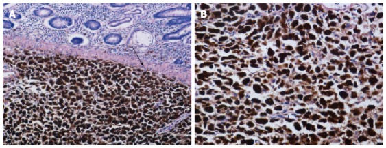
Figure 2.

Histopathologic findings of excised specimen. A: Microscopic examination showing tumor cells invading the colonic submucosa (arrow) (hematoxylin and eosin, × 100); B: Microscopic examination showing epithelioid and spindle tumor cells with melanin deposition in the cytoplasm (hematoxylin and eosin, × 200).
