
Figure 1.
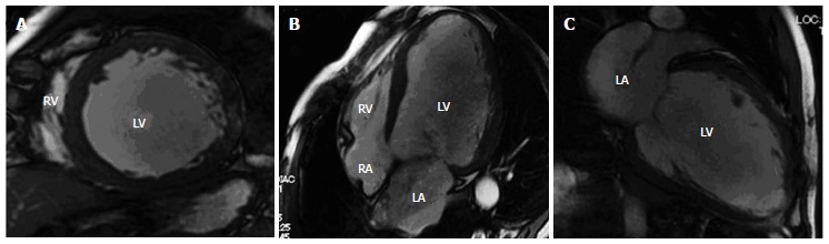
Representative cine-cardiac magnetic resonance images in a 62-year-old male patient with dilated cardiomyopathy. The images show mid-ventricular short axis (A), horizontal axis (4-chambers) (B) and vertical long axis views (C). The images reveal dilatation of left ventricular (LV) cavity and diffuse wall thinning (relatively homogenous). The LV end-diastolic volume, LV end-systolic volume, LV ejection fraction (EF) and LV mass are 329.1 mL, 252.5 mL, and 23.3%, 153.2 g, respectively. LV and RV: Left and right ventricles; LA and RA: Left and right atria; LV: Left ventricular.