


Figure 4.
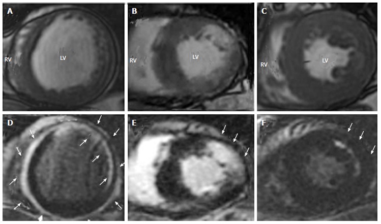
Representative cine-cardiac magnetic resonance (A-C) and late gadolinium enhancement-cardiac magnetic resonance (D-F) images in patients with cardiac sarcoidosis. A, D: A patient with LV dilatation, reduced LVEF (22%) and circumferential subepicardial and subendocardial LGE with spared mid-myocardium; B, E: A patient with reduced LVEF (38%) and nodular LGE in antero-lateral wall; C, F: A patient with preserved LVEF (58%) with mid-wall striated LGE in antero-lateral wall. White arrows indicate LGE areas. A part of the images is taken from Matoh et al[10] with permission. LGE: Late gadolinium enhancement; LV: Left ventricular; LVEF: Left ventricular ejection fraction