
Figure 7.
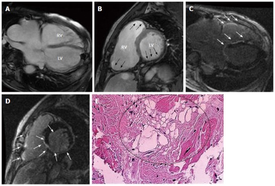
Representative cine- cardiac magnetic resonance (A, B) and late gadolinium enhancement-cardiac magnetic resonance (C, D) images in a 55- year-old male patient with arrhythmogenic right ventricular cardiomyopathy/dysplasia. The images show horizontal axis (4-chambers) (A, C) and mid-ventricular short axis (B, D) views. Cine-CMR images reveal dilatation of both RV and LV chamber. Focal dilatation of RV and wall thinning in inferior LV wall are also apparent (black arrows). LGE-CMR images show diffuse LGE in RV wall and in inferior LV wall (white arrows). A sub-endocardial biopsy demonstrates fatty infiltration in RV myocardium (Circle, H-E stain, 100×). LGE: Late gadolinium enhancement; LV: Left ventricular; LGE-CMR: Late gadolinium enhancement-cardiac magnetic resonance; RV: Right ventricular.