


Figure 8.
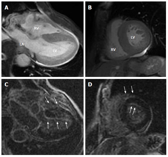
Representative cine-cardiac magnetic resonance (A, B) and Late gadolinium enhancement-cardiac magnetic resonance (C, D) images in a 76-year-old male patient with AL amyloidosis (IgAλ type multiple myeloma, Bence-Jones protein positive). The images show horizontal axis (4-chmbers) (A, C) and mid-ventricular short axis (B, D) views. Cine-CMR images reveal diffuse hypertrophy in LV and RV wall. LGE-CMR images show a characteristic subendocardial enhancement of the LV and RV with an atypically dark appearance of the blood pool (white arrows). LGE: Late gadolinium enhancement; LV: Left ventricular; LGE-CMR: Late gadolinium enhancement-cardiac magnetic resonance; RV: Right ventricular.