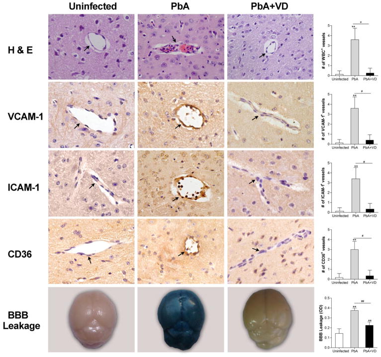
FIGURE 2.
VD treatment decreased endothelium activation and improved BBB integrity. On day 5 p.i., five mice from each of the three groups were processed for histology with H & E staining and immunohistochemical analysis with anti-ICAM-1, -VCAM-1, and -CD36 antibodies. Top four panels: Images show representative brain sections with the microvessels (arrows), while the corresponding bar graphs indicate quantification of leukocytes-, ICAM-1-, VCAM-1-, and CD36-positive microvessels, respectively. Microvessels per microscopic field were quantified in 20 fields per mouse, and values are mean ± SEM from five mice in each group. Bottom panel: Representative brain images show the extent of vascular leakage using the Evans blue extravasation method, while the corresponding bar graph shows quantitative assessment of BBB leakage, expressed as the optical density (OD) of brain extracts at 630 nm (mean ± SEM, n=5). ** indicates significant difference at P<0.05 (t-test) between uninfected control group and PbA-infected group, while # and ## indicate significant difference between the PbA and PbA+VD groups at P=0.05 and P=0.01, respectively.

