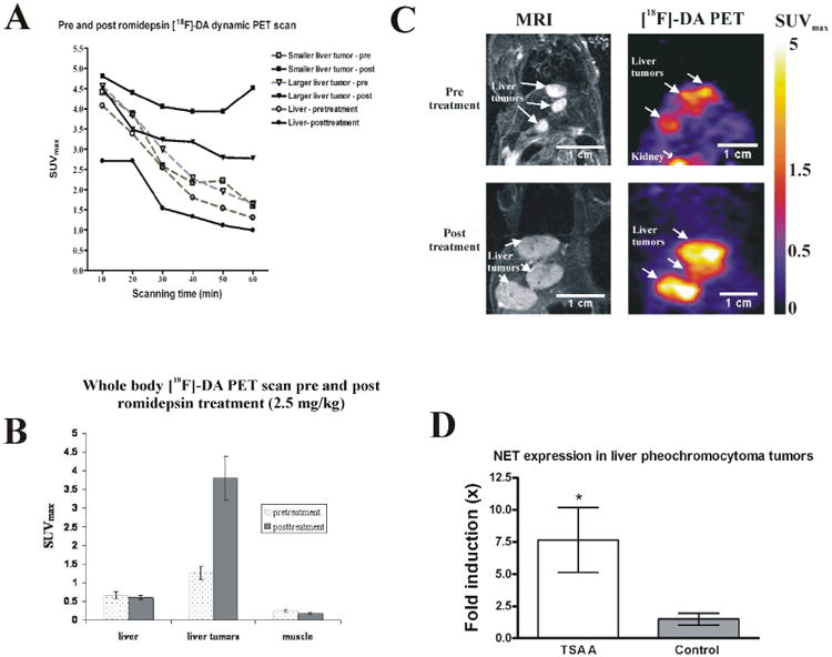
Figure 5.

(A) Dynamic PET acquisition captured the pharmacokinetics of [18F]-fluorodopamine in liver tumors, and in liver, both pre- and post-treatment, with a 60-minute imaging period. (B) PET imaging showing [18F]-fluorodopamine uptake as mean (±SEM) maximum standardized uptake values (SUVmax) 6 days before and 24 hr after treatment with a single intravenous dose of romidepsin (2.5mg/kg), in liver, pheochromocytoma liver metastases, and muscle. [18F]-fluorodopamine uptake in the metastatic liver lesions is significantly increased after administration of romidepsin compared to the uptake in the same lesions one week earlier on the pretreatment scan (p < 0.001). (C) Representative pre- and post-treatment PET/MRI images of the same mouse. There was one week between pre- and post-treatment scans. Tumor growth over that one week is presented in MRI (left panels), and the corresponding PET of [18F]-fluorodopamine uptake (right panel) was imaged 24 hours after MRI. (D) Relative mRNA expression of the norepinephrine transporter in liver lesion samples after trichostatin A treatment (n = 7) is higher than in control, untreated liver lesions (n = 6). RNA concentrations were normalized to beta-actin.
