
Figure 3.
Human PBMC-Derived iPSCs Give Rise to Functional CMs
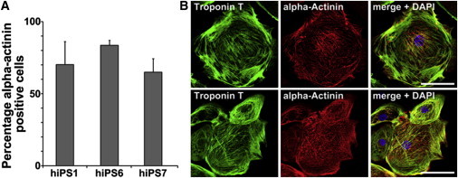
(A) High percentage of α-actinin-positive cells. After differentiation, cells were stained for the cardiac marker protein α-actinin and analyzed by a flow cytometer. Depending on the hiPSC line, up to 87% of the cells were positive for α-actinin (line hB53 hiPS6). Error bars, SD (n = 3 independent experiments).
(B) Representative images of troponin T- and α-actinin-stained hiPSC-derived CMs. Scale bars, 10 μm (upper panel) and 30 μm (lower panel).