
Figure 6.
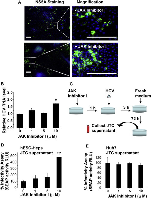
JAK Inhibitor I Improves HCV Infectivity in hESC-Heps
(A) hESC-Heps pretreated (bottom panel) or not (top panel) with 10 μM JAK inhibitor I were infected with HCVcc. At 3 days postinfection, cells were immunostained for viral NS5A antigen (green) and counterstained with DAPI (blue). Scale bar, 200 μm.
(B) hESC-Heps were pretreated with 0, 1, 5, or 10 μM JAK inhibitor I before infection. HCV RNA levels in infected hESC-Heps were measured by qPCR. The results are presented as fold change of HCV RNA level relative to that of mock-treated cells.
(C) Generation of JAK-inhibitor-I-treated conditioned medium (JTC). hESC-Heps or Huh7 were pretreated for 1 hr with 0, 1, 5, and 10 μM JAK inhibitor I, respectively, before infection with HCVcc and the JTC at 72 hr postinfection was collected.
(D) JTC of hESC-Heps improved HCV infectivity in human hepatoma cells. Huh7-J20 reporter cells were infected in advance by HCV for 3 hr, washed with PBS, and then incubated with JTC of hESC-Heps for 72 hr. The virus infectivity levels were determined by measuring SEAP activity in the medium.
(E) JTC of Huh7 has no effect on HCV infectivity in human hepatoma cells. Huh7-J20 reporter cells were infected in advance by HCV for 3 hr, washed with PBS, and then incubated with Huh7 JTC for 72 hr. The virus infectivity was determined as described in (D).
∗p < 0.05, ∗∗∗p < 0.001 compared with the group treated with 0 μM JAK inhibitor I. Error bars represent the SD of the mean. n = 3, biological replicates.