
Figure 3. hESC-Derived NC Cells Form DRG-Like Clusters.
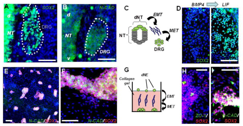
(A–C) Migratory NC cells reacquire both SOX2 (A) and N-CAD expression (B) after coalescing into embryonic day 10 (E10) dorsal root ganglia (DRG); (d) and (v) denote the dorsal-ventral orientation of the neural tube schematically illustrated (C).
(D) dNE cells reacquire SOX2 expression following LIF treatment. Left panel shows SOX2 staining in cells treated for 4 days with 100 ng/ml BMP4. 6.69 ± 1.25% of total cells were SOX2+. Four days of treatment with 100 ng/ml LIF (right panel) resulting in formation of numerous SOX2+ clusters with 34.15 ± 3.1% of total cells expressing SOX2.
(E and F) hES-derived migratory neural crest cells reform clusters after migration through the collagen gel; low (E) and high (F) magnifications.
(G) Schematic of human dNE migrating through collagen to form colonies at the bottom of the plate.
(H and I) Clusters of cultured dNE cells. SOX2+ cells are found in the polarized epithelial rosettes identified by the apical accumulation of clusters positive for the tight junction protein ZO-1 (H) and N-CAD (I). Scale bar, 100 mm; blue, Hoechst dye. Error bars ± SE.